Laser Scanning Microscope Facts

What Is Confocal Laser Scanning Microscopy

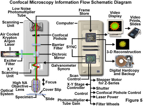
Olympus Fluoview Resource Center Theory Of Confocal Microscopy

What Is Confocal Microscopy Cherry Biotech

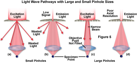
Rp Photonics Encyclopedia Confocal Scanning Microscopes Operation Principle Pinhole Fluorescence Microscopy

Confocal Laser Scanning Microscopy An Overview Sciencedirect Topics
Zeiss Microscopy Online Campus Introduction To Superresolution Microscopy

Fluorescent microscopy not only makes our images look good it also allows us to gain a better understanding of cells structures and tissue.
Laser scanning microscope facts.

Confocal Microscopy Microscopist Co Uk
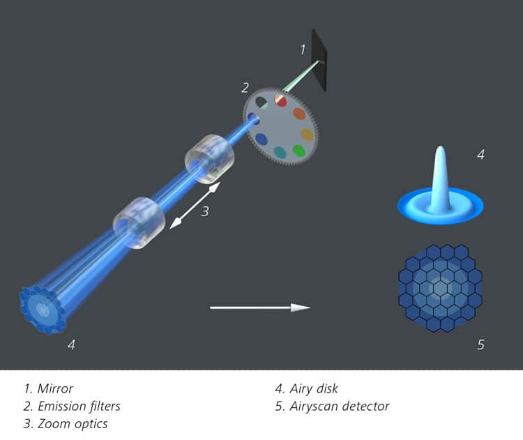
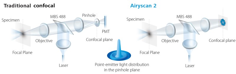
Zeiss Airyscan A Brave New Microscopy World With Sharper Confocal Resolution Bitesize Bio
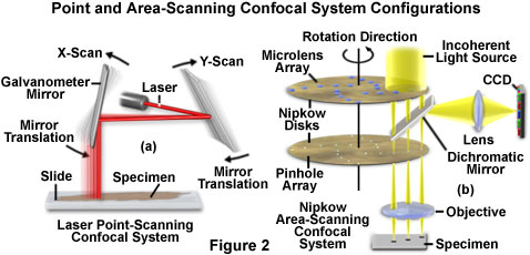
Confocal Microscopy Confocal Microscope Scanning Systems Olympus Life Science
Confocal Microscopy Introduction Olympus Life Science

Confocal Microscopy An Overview Sciencedirect Topics
Microscope Types Parts History Diagram Facts Britannica

Zeiss Microscopy Online Campus Introduction To Spinning Disk Microscopy

Confocal Microscopy Introduction And Applications Axiom Optics

Pin By Unity Smsc On Nature Tardigrade Microscopic Photography Micro Photography
Zeiss Microscopy Online Campus Digital Imaging Considerations

Scanning Electron Microscope Images Reveal Hidden Horror And Beauty Microscopic Photography Microscopic Images Scanning Electron Microscope
Application Note Multiplex Mode For The Lsm 9 Series With Airyscan 2 Fast And Gentle Confocal Super Resolution In Large Volumes

Photographer And Neurobiologist Igor Siwanowicz Captures The Striking Complexity Of Insects Using A Laser Sca Microscopic Photography Microscopic Insect Photos

Nikon Microscopyu Featured Microscopist Stephen W Paddock Biology Experiments Nurse Art Science Art

Interactions Between Soluble Dietary Fibers And Wheat Gluten In Dough Studied By Confocal Laser Scanning Microscopy Sciencedirect

Confocal Microscopy An Overview Sciencedirect Topics

The Extraordinary Details Of Tiny Creatures Captured With A Laser Scanning Microscope By Igor Siwanowicz Microscopic Microscopic Photography Macro And Micro

Neurolove Confocal Microscopy Image Things Under A Microscope
Https Encrypted Tbn0 Gstatic Com Images Q Tbn 3aand9gcr3fysoxor5w4y0kayjtt5nby84 Yhi3vdxn3rx2 E Usqp Cau
The Applications And Practical Uses Of Scanning Electron Microscopes Ata Scientific

Under The Microscope Some Things Look Too Crazy To Be Real Insects Insect Art Microscopic Photography

Confocal Laser Scanning Microscopy And Scanning Electron Microscopy Of Tissue Ti Implant Interfaces Sciencedirect
Https Nptel Ac In Content Storage2 Courses 102103044 Pdf Mod3 Pdf
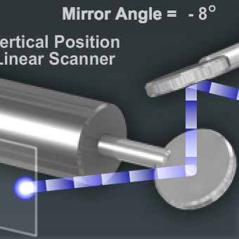
Resonant Scanning In Laser Confocal Microscopy Nikon S Microscopyu

Electron Microscope Microscopic Microscopic Photography Scanning Electron Microscope

Invisible Details Of Tiny Creatures Uncovered With Laser Microscope Photos Nikon Small World Microscopic Photography Microscopic Images

Bone Cells Imaged Using Confocal Microscopy Confocal Microscopy Microscopic Photography Microscopy Art
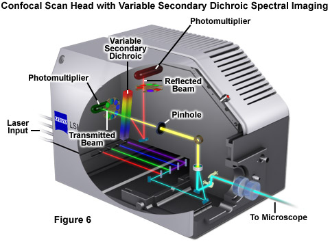
Zeiss Microscopy Online Campus Introduction To Spectral Imaging

800x Mikroskop Altinda Sivrisinek Ayagi Things Under A Microscope Microscopic Mosquito

Caffeine Under A Microscope Microscopic Photography Scanning Electron Microscope Science Images
Fig 5 A Scheme Of Principle For Laser Scanning Confocal Microscopy Two Galvanometer Mirrors Oscillating On Orthogon Confocal Microscopy Microscopy Schemes

Photographer And Neurobiologist Igor Siwanowicz Captures The Striking Complexity Of Insects U Microscopic Photography Macro Photography Insects Creature Design

Expose The Light Science And Photography The Radiolab Microscopic Photography Science Images Scanning Electron Microscope
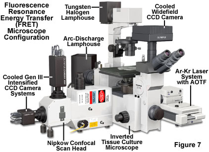
Fluorescence Resonance Energy Transfer Fret Microscopy Introductory Concepts Olympus Life Science

Confocal Laser Scanning Microscopy An Overview Sciencedirect Topics

The Race To Super Resolution Microscopy Is Deconvolution Enough Olympus Life Science

Tiniest Creatures Captured Using Laser Scanning Microscope With Images Microscopic Photography Jellyfish Art Photography

A High Performance Cost Effective Open Source Microscope For Scanning Two Photon Microscopy That Is Modular And Readi Laboratory Idea Open Source Microscopy

Pin On Microscope Images Facts History

What Is Circuit Optogenetics Biocompare The Buyer S Guide For Life Scientists

Confocal Microscopy Laboratory Nus Centre For Bioimaging Sciences Cbis
Laser Safety Nikon S Microscopyu

Confocal Microscopy Spectral Bleed Through Artifacts In Confocal Microscopy Olympus Life Science
Https Encrypted Tbn0 Gstatic Com Images Q Tbn 3aand9gcs8sytwgmkmgf6fmhtx9v0yupwtla Tc8ut 6vwtnyf131bovul Usqp Cau
Source : pinterest.com
